Click for pdf: Clearing the C-spine
Introduction:
Injury to the cervical spine is rare in children and is most often seen in cases of blunt trauma. It is essential that when one suspects cervical injury, appropriate evaluation and management must be undertaken. Clearance of the cervical spine is a method that is used to prevent the possibility of quadriplegia and complications of immobilization. Spinal trauma results from four main mechanisms consisting of hyperflexion, hyperextension, axial loading and lateral rotation. Children 8 years of age or younger, tend to have more upper cervical injuries when compared with adults because of the anatomy of their bodies.
In order to understand the critical aspects of cervical spine injury, it is important to acknowledge the normal parameters of the pediatric cervical spine described in table 1.
Table 1. Normal Parameters of the Pediatric Cervical Spine
| Parameter | Normal Value |
| C-1 facet-occipital condyle distance | ≤ 5 mm |
| Atlanto-dens interval | ≤ 4 mm |
| Pseudosubluxation of C2 on C3 | ≤ 4 mm |
| Pseudosubluxation of C3 on C4 | ≤ 3 mm |
| Retropharyngeal space | ≤ 8 mm (at C-2) |
| Retrotracheal space | ≤ 14 mm (at C-6, under age 15 yr.) |
| Tong ratio (canal to vertebral body) | ≥ 0.8 |
| Space available for cord | ≥ 14 mm |
Table 1. Adapted from Tornetta and Einhorn, 2004.
Clinical Presentation
The pediatric cervical spine does not become adult like until about the age of 8 years. The unique anatomic features of children aged 8 years of age or younger are described in table 2. Considering the large difference in ratio of head size to body the infant will experience different inertial forces compared to fully matured individuals. Ligaments and joint capsules are more lax, facets are more horizontal, and the vertebral bodies are wedge shaped. These factors increase the risk of injury to the levels of C1 and C2.
Cervical spine injuries occur mainly in the upper cervical spine above C4 in patients 8 years of age or younger which most often involve the occiput, C1, and C2 complex and thus carries increased risk of fatality. Patients older than 8 years of age typically sustain more injuries below C4 and carry a much lower fatality rate. Up to 30% of traumatic spine injuries in children present as a traumatic myelopathy known as spinal cord injury without radiographic abnormality (SCIWORA).
Unique Anatomic Features of Children Age 8 and Younger
- Large head-to-body ratio
- Ligamentous laxity
- Relative paraspinal muscle weakness
- Horizontal, shallow facet joints
- Increased spinal column elasticity
- Forces dissipated over several adjacent segments
- SCIWORA possible
- Presence of the ring apophysis
- Fractures traverse vertebral body growth plate.
Adapted from Tornetta and Einhorn (2004).
History:
Although it is not an easy task, thehistory can be very useful. Elements of the history that are critical include the cause of trauma, the mechanism of injury and the presence of any symptoms at the time of injury. . A spinal cord injury should be suspected if the child has a history of numbness, tingling, or brief paralysis.
Some children are predisposed to cervical injuries more than others and this should be taken into consideration when taking a history. This includes children with Down Syndrome (atlano-axial instability), Klippel-Feil syndrome (congenital fusion of cervical spine), previous cervical spine surgery, and other syndromes affecting the cervical spine.
The major causes of cervical spine injury include trauma associated with severe force, diving, acceleration-deceleration injury and severe multisystem trauma. The mechanism on injury may also predict the type of injury to expect. Hyperflexion injuries are most common and are associated with wedge fractures of the anterior cervical bodies with disruption of posterior aspects.
The history must include the presence of symptoms even if they have already resolved at the time of evaluation. The classic triad of symptoms of cervical spine injury is localized neck tenderness, muscle spasm and decreased range of motion of the neck. Neurologic symptoms may also be present such as burning, weakness, and dysthesias. Some patients may present without any symptoms but this does not exclude spinal injury.
**Any child with symptoms suggestive of cervical spinal injury should be immediately immobilized and sent for radiographic evaluation**.
Physical Examination:
Physical examination should include vital signs, examination of the neck and neurologic examination. The skin should be inspected for any visible evidence of spinal traumancluding abrasions, edema, or ecchymosis. Pain or step-off along the spinous processes should also raise suspicion. Range of motion should only be attempted when the child is conscious and cooperative and an unstable injury is not suspected.
The National Emergency x-radiography utilization study (NEXUS) has developed a list of characteristics that are used to evaluate a patient as having a low probability of injury if they meet all of the following five criteria:
- No midline cervical tenderness
- No focal neurological deficit
- Normal alertness
- No intoxication
- No painful, distracting injury
Patients with impaired consciousness:
Neurological examinations are limited in patients who are consciously impaired and can be very challenging for professionals to diagnose. However, as much of the neurological examination as possible should be performed as long as safety of the patient is maintained. The main goal while diagnosing consciously impaired patients is to prevent further damage to neural structures. If a child is likely to remain unconscious for a prolonged period of time (>48 hours) an MRI scan is recommended to diagnose any possibilities of a significant injury.
Diagnosis: Trauma Evaluation
All unconscious patients with an injury above the clavicles and those involved in high speed vehicular accidents should be assumed to have a spinal injury. Table 3, adapted from Kaji and Hockberger (2007) describes the factors in which suspected cervical spine injury is plausible:
Suspect C-spine Injury Based Upon
History:
- Mechanism of injury
- Motor vehicle crash (vs. vehicle, pedestrian or bicycle
- Falls > 4 feet or > 5 steps
- Driving or tackling incident
- Severe blunt trauma to head, neck or upper body
- Pre-existing conditions
- Previous neck surgery
Physical Assessment:
- Neck pain, tenderness or stiffness in awake and alert patient
- Altered level of consciousness (unconscious, altered mental status, GCS < 14)
- Abnormal peripheral motor sensory exam
- Altered vital signs
- Presence of other injuries (skull fx, facial and/or upper body injury
Early and proper immobilization in all patients with suspected cervical spine injury is crucial to avoid any further damage to the spinal cord. Spine board immobilization can be administered to avoid propagating further injury. This can be done with a spine board with an occipital recess or by placing a mattress or blankets beneath the shoulders and trunk of the child. A screening cross-table lateral x-ray of the spine, in addition to antero-posterior pelvis and chest x-rays, should be standard in the evaluation of all trauma patients.
In addition to proper spine board immobilization, a rigid cervical orthosis, properly designed for infants or children, should be applied. Sandbags on each side of the head will prevent motion. Until the cervical spine is cleared movement of the patient should only be performed with in-line traction using a log-roll technique.
Investigations:
Radiographic analysis
If a cervical spine injury is suspected radiologic investigation must be performed. This should include 3 views: lateral, AP and open–mouth odontoid. Children with clinical findings consistent with spinal injury in the face of a normal radiograph should still be treated as if they have an injury. You must remember your ABCS when reading C-spine radiographs: Alignment, Bones, Cartilage and Soft Tissue.
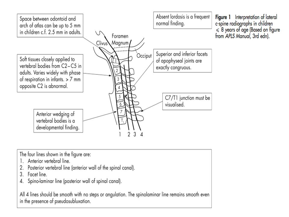
Lateral View: It is critical that all 7 cervical vertebrae and T1 be visualized in this view. Bony abnormalities such as fractures, subluxations, and dislocations can be seen on the lateral radiograph. You must also assess alignment, cartilaginous structures and soft tissue.
AP View: The spinous processes should be aligned in the midline on this view. It may identify lateral fractures not seen on the lateral view.
Open Mouth Odontoid View: This is the best view to visualized the odontoid process (dens) and the body of C2 in between C1 in children over 9 years of age. In younger children, Waters view may be used.
Flexion Extension View: This view may be useful if the other 3 views are negative. It may reveal instability of the cervical spine and ligamentous injuries.
It is also important to take into account the differences in the pediatric patient in regards to reviewing the cervical spine radiograph. Injuries to the spinal column can often be subtle and absent on initial radiographs. Successful treatment is based on knowledge of the radiographic, anatomic and developmental differences between the pediatric and adult spine. Table 4 describes these differences:
Differences in the Cervical Spine seen in Pediatric Patients
| Pseudosubluxation |
|
| Localized Kyphosis |
|
| Overriding C1 over Tip of Odontoid (C2) |
|
| Persistence of Basilar Odontoid Synchondrosis |
|
Adapted from Tornetta and Einhorn (2004).
Figure 1. Interpretation of lateral c-spine radiographs in children < 8 years of age. Adapted from Slack and Clancy (2004).
Evaluation
The cervical spine is evaluated as follows:
| Procedure | Evaluation Checklist |
| Lateral C-spine radiograph | Must see top of T1 |
| Assess alignment | Anterior vertebral linePosterior vertebral lineSpinolaminar lineSpinous process line |
| Check radiographic relationships | Atlanto-dens intervalRetropharyngeal and retro tracheal spacesSpace available for the cord |
| Antero-posterior and open mouth odontoid views | Alignment of lateral massesOdontoid fractureInterpedicular distance |
| Flexion-extension views | Assess ligamentous stabilityActive range of motion onlyPhysician supervisedFully cooperative patient
May need to wait until the first follow-up visit |
Adapted from Tornetta and Einhorn (2004).
In most children it may be practically impossible to obtain views of open mouth as there is much cooperation required in holding the mouth open; some professionals have recommended avoiding open mouth views in children less than 9 years of age.
If the C7-T1 junction cannot be viewed at the first attempt, the shoulders may be depressed by pulling on the arms, or oblique projections, or swimmer’s views may be taken. Repeated attempts at plain views should be avoided and if C7-T1 still cannot be visualized, CT is advised.
If there is a neurological deficit and/or ligamentous or soft tissue injury, MRI is the best procedure to perform; however, may only be suitable for stable patients.
Differential Diagnosis:
Developmental Features That Can Be Misinterpreted as Injury
| Radiographic Finding | Misinterpretation | Explanation |
| Absent cervical lordosis | Muscle spasm from C-spine injury | Seen in up to 14% of patients less than 8 yr. of age. |
| Dentocentral synchondrosis | Odontoid fracture | Usually not fused by age 6; lucency below level of body dens interface |
| Wedge-shaped vertebrae | Compression fracture | Usually present up to age 8; adjacent bodies similar |
| Incomplete ossification of ring apophysis (secondary ossification) | Avulsion fracture | Appear at age 5, fuse between age 18 and 25 |
| Notching of anterior and posterior vertebral body in infancy | Vertebral body fracture | Vascular channels. Anterior disappears at age 1. Posterior remains throughout life. |
Selected Congenital Vertebral Anomalies
| Anomaly | Characteristic | Radiographic significance |
| Os odontoideum | Rounded hypoplastitic apical segment of dens.Remnant of axis at base | Can be confused with dens fracture.May represent nonunion. May require fusion |
| Klippel-Feil syndrome | Congenital fusion of two or more vertebrae | Longer lever arm may lead to higher incidence of fractures |
| Down syndrome | Ligamentous laxity leads to decreased atlanto-dens interval / space available for the cord. | Atlanto-axial instability, myelopathy. |
Management
Atlanto-Occipital Dislocation
- Very unstable
- Requires halo immobilization if neurologically intact
- Fusion if instability remains or neurological deficit
- Occiput-C1 fusion if neurologically intact
- Occiput-C2 fusion if neurologically impaired
Atlas Fractures
- Halo immobilization or Minerva brace
- Up to 6 months of immobilization may be necessary
- Surgical intervention rarely indicated
Atlantoaxial (C1-C2) Disruptions
- Rotatory subluxation can be treated with temporary immobilization
- Rarely will require traction for reduction (older than 1 week)
- Dislocation or ligamentous instability less predictable
- Initial treatment with Halo immobilization for 8 to 12 weeks
- C1-C2 fusion if instability persistent
Hangman Fracture (Pedicle Fracture C2)
- Most respond to closed reduction and halo vest for 8 weeks
- Posterior C1-C3 fusion indicated for non-union or significant disc disruption
C2-C3 Subluxation and Dislocation
- Younger than 8 years, closed reduction and halo vest immobilization
- Older than 8 years, usually require posterior fusion
Middle to Lower Cervical Injuries
- Younger than 8 years, closed reduction and halo vest immobilization
- Initial trial of Halo immobilization for 3 months for ligamentous instability
- Posterior fusion if instability persists beyond 3 months
- Early surgical stabilization for unstable fractures or spinal cord injury
Conclusion
Clearance of the pediatric cervical spine can be a challenging diagnosis and requires great understanding of the pediatric spine. Although much of the principles are the same as for adults, certain aspects in regards to evaluation with radiography and anatomic features are important in proper diagnosis and treatment of children. Inadequate adherence to procedures in emergency situations can result in further injury and complications. A thorough history, physical exam and radiographic evaluation must be completed and proper immobilization if a cervical spinal injury is suspected.
References
Kaji, A., Hockberger, R. 2007. Imaging of Spinal Cord Injuries. Emergency Medicine
Clinics of North America, 25 (3): 735-750
Slack, S.E., Clancy, M.J. 2004. Clearing the cervical spine of pediatric trauma
patients. Journal of Emergency Medicine, 21: 189-193
Tornetta P., Einhorn T. Pediatrics. Lippincott Williams and Wilkins, 2004.
Caviness C. Evaluation of cervical spine injuries in children and adolescents.
Acknowledgements:
Written by John Hilhorst
Edited by Anne Marie Jekyll


 (3 votes, average: 4.33 out of 5)
My 10 yr old daughter has c2, c3 posterior fusion with pressure on her spinal cord from part of the disc(what is left of it ). I was wondering what can be done. She also has Fluid in and around C7, T1 this fluid also goes into the muscle.
She is developmently and congnitivly delayed with seizure disorder and migrane head aches. We take her to Denver Childrens hospital.